
Abstract
This study reports a case series of glaucoma patients in whom a biodegradable collagen-glycosaminoglycan implant (Ologen®) failed to prevent fibrosis after non-penetrating deep sclerectomy (NPDS) and describes the associated histopathological findings. This single-centre retrospective study included four patients with progressive glaucoma and inadequate intraocular pressure (IOP) control despite maximal medical therapy. All underwent NPDS with implantation of a collagen-glycosaminoglycan matrix and adjunctive mitomycin C. Postoperative follow-up included IOP monitoring, slit-lamp examination, and photographic documentation. Patients with bleb failure required surgical revision, and excised tissue, including the implant and surrounding Tenon’s capsule, was analysed histologically. All cases developed progressive bleb fibrosis. The implants remained intact, encapsulated by dense fibrotic tissue (“ring-of-steel” appearance), with minimal fibroblast infiltration, extensive collagen deposition, and no significant degradation for up to three years. These findings highlight variability in tissue response and suggest a potential role of patient-specific fibrotic predisposition in surgical outcomes.
Keywords: Biodegradable collagen-glycosaminoglycan implant, glaucoma surgery, fibrosis, wound modulation, biodegradation
Introduction
Filtering surgery remains the most widely used surgical intervention for glaucoma. These procedures aim to create a fistulous tract between the anterior chamber and the subconjunctival space to facilitate the drainage of aqueous humour and reduce intraocular pressure (IOP). As a result, aqueous humour accumulates in a subconjunctival reservoir known as the filtration bleb. The primary cause of surgical failure is excessive formation of fibrotic tissue around the new drainage pathway,, which compromises proper functioning of the bleb.
Glaucoma surgery is intrinsically pro-fibrotic due to a chronic pro-inflammatory conjunctival environment that often results from prolonged use of topical medications, preservative agents in eye drops, hypersensitivity to active compounds, and a high prevalence of dry eye disease. Furthermore, postoperative tissue exposure to aqueous humour, which is rich in pro-inflammatory cytokines, promotes fibroblast activation.
Fibrotic scarring involves the excessive synthesis of extracellular matrix (mainly consisting of dense, organised collagen fibres) and the persistence of myofibroblasts that should normally undergo apoptosis. Effectively modulating this fibrotic response is crucial for improving surgical outcomes.
Advances in tissue engineering have provided alternative strategies to modulate wound healing and reduce scar formation after glaucoma surgery, such as biodegradable implants. Unlike antimetabolites, whose non-specific inhibition of cell proliferation may lead to significant ocular toxicity, these implants promote the formation of functionally effective filtering blebs while degrading into non-toxic byproducts, thereby eliminating the need for surgical removal.
Ologen® (Aeon Astron Europe B.V., Leiden, the Netherlands) is a biocompatible, biodegradable implant composed of collagen (90%) and glycosaminoglycan (10%)., These components are organised in a three-dimensional system, creating a 95% porous structure that acts as a biological scaffold for the healing process,, , facilitating fibroblast growth inside the implant and guiding their migration to create a loose, randomly organised deposition of connective tissue similar to physiological conditions., , , This is the mechanism underlying the implant’s modulation of wound healing., , , However, it is important to highlight that the implant itself does not have antifibrotic properties and could therefore hypothetically cause a secondary tissue reaction in the subconjunctival space. To avoid this reaction and modulate its immunogenicity, the collagen is purified with pepsins that eliminate telopeptides, which are the N- and C-terminal amino acid sequences responsible for the antigenicity of the collagen molecule., , ,
Various published surgical series comparing patients undergoing surgery with and without the implant have yielded inconclusive results regarding its potential to enhance the antifibrotic effect of other techniques (summarised in , , , , - and 2, , , , -). In this case series, we present four cases of glaucoma surgery in which this biodegradable collagen-glycosaminoglycan implant failed to prevent fibrosis and was associated with an encapsulating fibrotic response surrounding the implant, which ultimately compromised surgical success.
Table 1. Published studies evaluating the safety and efficacy of Ologen® and MMC as adjuvants in trabeculectomy surgery for glaucoma
- | Study | Type of study | Groups | N | Follow-up (months) | Outcomes | MMC dose | Complications | ||||||||||
|---|---|---|---|---|---|---|---|---|---|---|---|---|---|---|---|---|---|---|
Compared to MMC-enhanced trabeculectomy | OLO > MMC | Yuan et al. 2015 | Prospective randomised | Trab + OLO | 31 | 60 | Higher success rates (complete and qualified) in OLO group Lower IOP in OLO group | 0.2 mg/mL 2 min | No significant differences between groups | |||||||||
Trab + MMC | 32 | |||||||||||||||||
El-Sayyad et al. 2017 | Prospective randomised (JOAG) | Trab + OLO | 20 | 12 | Lower IOP at end point in OLO group Better MBGS scoring of blebs in OLO group | 0.4 mg/mL 2 min | Higher rate of hypotony in MMC group Less needling and suture lysis in OLO | |||||||||||
Trab + MMC | 20 | |||||||||||||||||
OLO = MMC | Cillino et al. 2011 | Prospective randomised | Trab + OLO | 20 | 24 | No differences in mean IOP and IOP reduction at end point. No differences in complete and qualified success at any target IOP Higher bleb height in OLO group | 0.2 mg/mL 2 min | Higher incidence of early hypotony in MMC, although not statistically significant Higher incidence of early bleb leak in OLO group | ||||||||||
Trab + MMC | 20 | |||||||||||||||||
Chelerkar et al. 2021 | Prospective non-randomised | Phaco + Trab + OLO | 40 | 12 | No statistically significant differences in IOP or bleb height, area, and vascularity | 0.2 mg/mL 2 min | No significant differences between groups | |||||||||||
Phaco + Trab + MMC | 53 | |||||||||||||||||
Glandorf et al. 2020 | Retrospective | Trab + MMC | 43 | 12 | No differences in IOP reduction, complications, or need for additional medical treatment Greater success with MMC alone, although not statistically significant | 0.2 mg/mL 1 min | No significant differences between groups | |||||||||||
Trab + MMC + OLO + BVZ | 46 | |||||||||||||||||
Trab + MMC + OLO + intracameral BVZ | 41 | |||||||||||||||||
Paul et al. 2022 | Prospective non-randomised | Phaco + Trab + OLO | 18 | 12 | No differences in IOP reduction or need for additional medical treatment Better bleb morphology in OLO group | 0.2 mg/mL 1 min | No significant differences between groups | |||||||||||
Phaco + Trab + MMC | 16 | |||||||||||||||||
Navero-Rodríguez et al. 2024 | Retrospective non-randomised | Trab + MMC | 44 | 36 | No differences in IOP reduction, use of topical medications, or complete and qualified success rates More suture release and bleb needling with MMC alone | 0.2 mg/mL 2 min | No significant differences between groups | |||||||||||
Trab + OLO | 34 | |||||||||||||||||
Trab + OLO + MMC | 41 | |||||||||||||||||
Compared to MMC-enhanced trabeculectomy | OLO < MMC | Rosentreter et al. 2010 | Prospective randomised | Trab + OLO | 10 | 12 | Lower IOP at end point in MMC group Higher complete success rate in MMC group More avascularity in MMC group | 0.2 mg/mL 3 min | No significant differences between groups | |||||||||
Trab + MMC | 10 | |||||||||||||||||
Senthil et al. 2013 | Prospective randomised | Trab + OLO | 19 | 24 | Lower mean IOP reduction at 6 months in OLO group No differences at 12 and 24 months | 0.4 mg/mL 2 min | More hyphaema in OLO group | |||||||||||
Trab + MMC | 20 | |||||||||||||||||
Rosentreter et al. 2014 | Prospective randomised | Trab + OLO | 15 | 12 | Lower IOP at end point in MMC group Higher success rate in MMC group | 0.2 mg/mL 3 min | No significant differences between groups | |||||||||||
Trab + MMC | 15 | |||||||||||||||||
Narayanaswamy et al. 2013 | Prospective non-randomised | Phaco + Trab + OLO | 33 | 12 | Lower IOP in MMC group Greater bleb area in MMC group OLO blebs were more vascular Needling required more frequently in OLO group | 0.4 mg/mL 2 min | No significant differences between groups | |||||||||||
Phaco + Trab + MMC | 33 | |||||||||||||||||
Sen et al. 2018 | Prospective randomised | Trab + MMC | 25 | 12 | Greater IOP reduction in MMC group Blebs more diffuse in MMC group More avascular blebs in MMC group | 0.1 mg/mL 1 min | More hypotony in MMC group | |||||||||||
Trab + MMC + OLO | 25 | |||||||||||||||||
Compared to standalone Trab | Papaconstantinou et al. 2010 | Prospective randomised | Trab + OLO | 20 | 6 | No significant differences between groups in IOP or topical medication use | - | No significant differences between groups | ||||||||||
Trab standalone | 20 | |||||||||||||||||
Compared to standalone and enhanced Trab | Khairy et al. 2023 | Prospective randomised (PCG) | CTT without augmentation | 21 | 12 | No differences in IOP at end point No differences in success rates, failure rates, or survival rates between groups | 0.2 mg/mL 3 min | No significant differences between groups No report of hypotony or shallow AC in OLO group | ||||||||||
CTT + MMC | 21 | |||||||||||||||||
CTT + OLO | 21 | |||||||||||||||||
Non-comparative | Angmo et al. 2017 | Retrospective | Trab + OLO + MMC | 23 | 24 | Significant reduction in IOP and use of hypotensive medications Complete success in 92.6% Failure in 3.7% | 0.1 mg/mL 1 min | Shallow AC in 14.8% Encapsulated blebs requiring needling in 11.1% | ||||||||||
Dada et al. 2013 | Retrospective | Trab + OLO+MMC | 33 | 12 | Significant reduction in IOP and use of hypotensive medications | 0.1 mg/mL 1 min | 2 cases of shallow AC 2 cases of encapsulated blebs requiring needling | |||||||||||
MMC: Mitomycin C, OLO: Ologen, Trab: Trabeculectomy, IOP: Intraocular pressure, JOAG: Juvenile open-angle glaucoma, MBGS: Moorfields Bleb Grading System, CTT: Combined trabeculotomy and trabeculectomy, Phaco: Phacoemulsification, BVZ: Bevacizumab, PCG: Primary congenital glaucoma, AC: Anterior chamber
Case Reports
Case 1
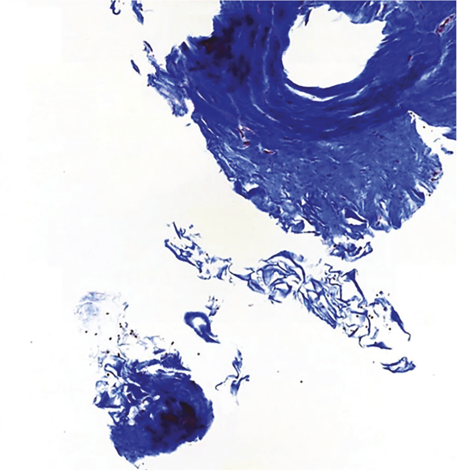
A 56-year-old female patient with moderate primary open-angle glaucoma (POAG) underwent non-penetrating deep sclerectomy (NPDS) with an intrascleral Esnoper V Clip implant, subconjunctival Ologen® implantation (6 mm×2 mm), and a subconjunctival injection of 0.1 mL 0.01% mitomycin C (MMC). The patient exhibited progressive bleb flattening during the postoperative follow-up, causing a rise in IOP and requiring additional hypotensive medical therapy. One year after the initial surgery, the patient underwent surgical bleb revision. Intraoperatively, fibrotic conjunctiva and Tenon’s capsule were observed encasing the intact Ologen® implant. Histopathological analysis revealed focal areas of dense fibrosis within the collagen matrix (), with disorganised collagen bundles and scattered inflammatory cell infiltration, indicating a chronic foreign body reaction. Additionally, no significant degradation of the implant was observed.
Case 2
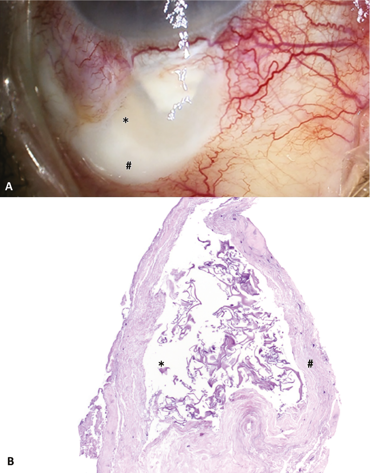
An 85-year-old female patient diagnosed with advanced POAG presented with inadequately controlled IOP despite maximally tolerated topical treatment. She underwent NPDS with a subconjunctival MMC injection and Ologen® implantation (6 mm×2 mm). During the postoperative course, the bleb developed an avascular appearance with cystic spaces and fluorescein leakage (). The patient was therefore scheduled for surgical bleb revision. Intraoperatively, the Ologen® implant was found to be intact 18 months after implantation, surrounded by a dense fibrotic capsule resembling a “ring-of-steel” reaction. This caused retraction of Tenon’s capsule, leaving an area of thin, avascular limbal conjunctiva. Histopathological analysis () confirmed the absence of Ologen® degradation, minimal fibroblast migration into the scaffold, and a thick wall of organised collagen deposition surrounding the implant.
Case 3
A 56-year-old female patient with a history of diabetic retinopathy was referred for primary angle-closure glaucoma. The patient developed toxicity associated with hypotensive treatment, and glaucoma progression was noted in the left eye. Therefore, NPDS with MMC injection and Ologen® implantation (6 mm×2 mm) was performed. Postoperatively, the bleb progressively became fibrotic, necessitating an escalation in hypotensive treatment. Bleb revision was performed three years after the initial procedure. Intraoperative findings revealed a highly retracted Tenon’s capsule that had contracted the bleb around the Ologen® matrix, which had remained intact for three years (). Histopathological assessment
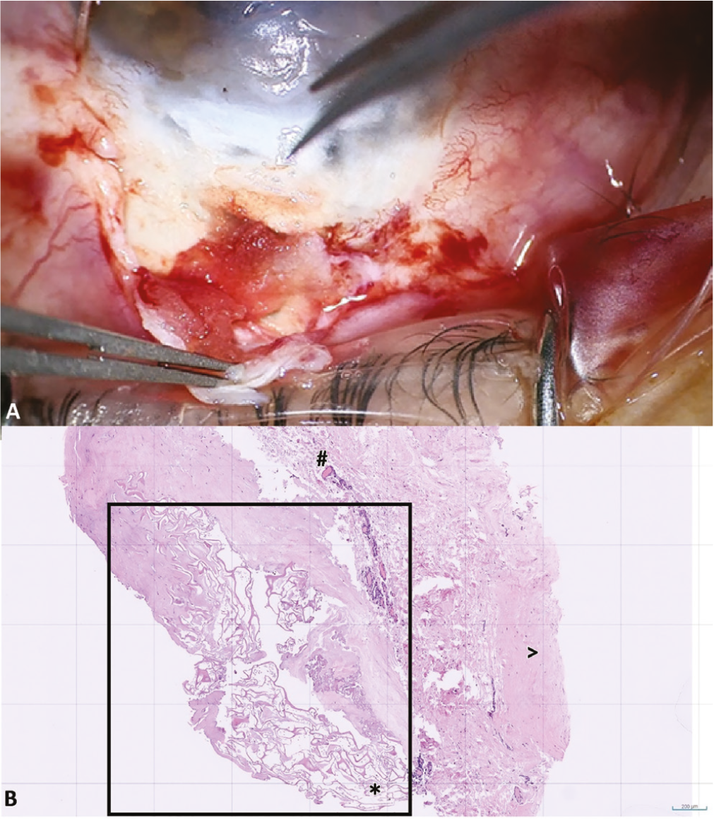
() revealed dense fibrotic tissue surrounding exogenous mesh-like material (Ologen®).
Case 4
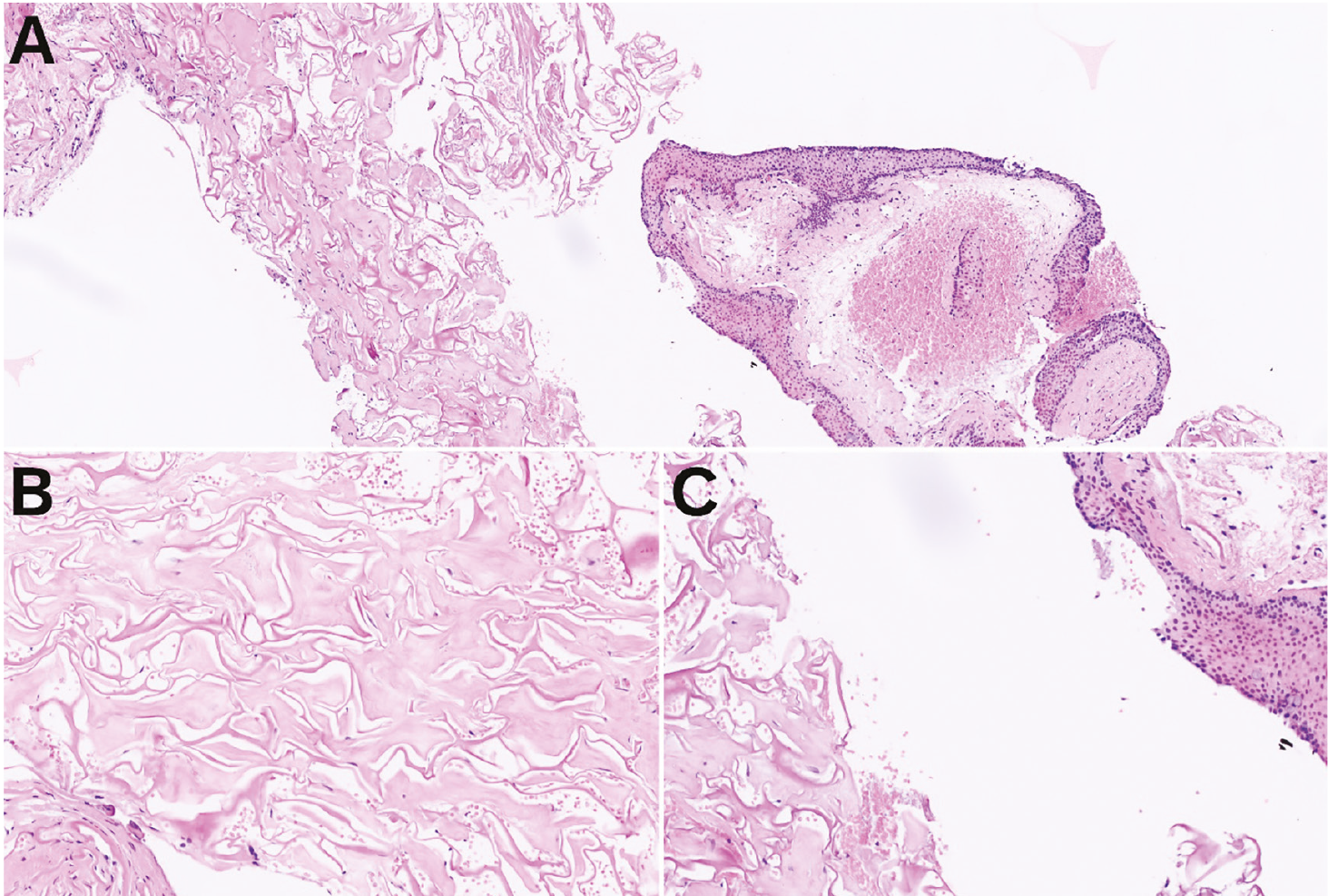
A 53-year-old female patient diagnosed with pigmentary glaucoma presented with a long history of topical antiglaucoma therapy. Visual field progression warranted a combined phacoemulsification and NPDS procedure enhanced with MMC and Ologen® implantation (6 mm×2 mm). IOP control remained inadequate a year after the procedure, with a fibrotic bleb that required needling on two separate occasions to reach target IOP. During surgical bleb revision, an intense scarring reaction was observed around the scleral flap and Ologen® implant. The implant was encapsulated in a manner similar to a foreign body reaction. After debridement of the fibrous tissue and removal of the residual Ologen® matrix, a single-layer pericardial graft was placed to reinforce the scleral flap. Histological examination revealed the Ologen® material embedded within a hyalinised collagenous matrix with minimal fibroblast presence ().
Following bleb revision, all patients demonstrated improved bleb function and better IOP control. No additional surgical procedures were required during follow-up, and IOP remained stable with or without adjunctive medical therapy depending on the case.
Discussion
Numerous studies have examined the effectiveness of Ologen® across several types of glaucoma surgery (summarised in , , , , - and , , -). However, interpretation is challenging due to variability in study design, limited sample sizes, and differences in surgical technique, including MMC timing and dosage. The heterogeneity of methodologies, from randomised trials to retrospective studies, further limits generalisability, resulting in inconsistent and sometimes contradictory findings.
Table 2. Studies evaluating the safety and efficacy profile of Ologen® in other types of glaucoma surgery
Surgery | Study | Type of study | Groups | N | Follow-up (months) | Outcomes | MMC dose | Complications | |||
|---|---|---|---|---|---|---|---|---|---|---|---|
GDD | Rosentreter et al. 2010 | Retrospective | Excision of GDD capsule + MMC + OLO | 10 | 8-12 | Adding OLO to capsule excision + MMC yielded higher success rate when revising GDD No significant differences in IOP | 0.2 mg/mL 2 min | - | |||
Excision of GDD capsule + MMC | 9 | ||||||||||
Salimi et al. 2019 | Retrospective | Excision of GDD capsule + MMC + OLO | 12 | 36 | No significant differences between groups in IOP reduction, hypotensive medications or success rate Valid strategy for GDD revision, successful in reducing IOP in both groups | 0.5 mg/mL 2 min | Significantly lower rate of complications in OLO group – no report of erosion of GDD, although MMC group had an erosion rate of 25% | ||||
Excision of GDD capsule + MMC | 11 | ||||||||||
Song et al. 2017 | Retrospective | Conventional GDD | 24 | 6 | Same IOP reduction, no differences in success. Enzymatic degradation rate of OLO-7 was slower | - | Lower hypotony rates in OLO groups than in conventional surgery, and less hyphaema or choroidal effusion. | ||||
GDD + OLO-6 | 20 | ||||||||||
GDD + OLO-7 | 24 | ||||||||||
Stephens et al. 2016 | Retrospective | Report of erosion rates when using OLO over GDD tube | 43 | 12-45 | 95.4% required no intervention for tube erosion | - | - | ||||
Kim et al. 2018 | Retrospective | GDD + OLO over plate | 8 | 12 | No differences between groups in overall IOP reduction, complete or qualified success. Number of IOP-lowering medications was significantly lower in OLO group during the early hypertensive phase | - | No differences in complication rates. | ||||
Conventional GDD | 12 | ||||||||||
Sastre-Ibáñez et al. 2019 | Prospective randomised | GDD + OLO over plate | 27 | 12 | No differences between groups in IOP reduction, complete or qualified success or survival rates. | - | No differences in complication rates. | ||||
Conventional GDD | 31 | ||||||||||
Suzuki Junior et al. 2023 | Retrospective multicentre case-control | GDD + OLO over plate | 25 | 6 | No significant differences between groups in IOP reduction, hypotensive medication use, or complete success rate Better outcomes in OLO group for qualified success | - | No differences in complication rates. | ||||
Conventional GDD | 100 | ||||||||||
NPDS | Guedes et al. 2019 | Retrospective | Phaco-NPDS + MMC + OLO | 51 | 12 | Significantly lower IOP and greater IOP reduction in OLO group Lower rate of medication use in OLO group | 0.2 mg/mL subconjunctival injection | Higher rate of needling in group without OLO | |||
Phaco-NPDS + MMC | 49 | ||||||||||
Aptel et al. 2009 | Prospective | NPDS enhanced with OLO in scleral bed | 15 | 3 | Significant reduction in IOP Large fluid-filled spaces on AS-OCT, lower reflectivity in OLO group | - | - | ||||
Elmekawey et al. 2020 | Retrospective | NPDS + OLO | 20 | 6 | Significant reduction in IOP and better bleb morphology in OLO group | - | - | ||||
NPDS | 20 | ||||||||||
XEN | Park et al. 2022 | Retrospective | XEN (± Phaco) + OLO | 61 | 6 | No significant differences in IOP reduction or antiglaucoma medication | 0.4 mg/mL 2 min | Hypotony reported in OLO-augmented group, possibly related to increased filtration facilitated by aqueous humour absorption by OLO No significant differences between groups | |||
XEN (± Phaco) | 42 | ||||||||||
Navero-Rodríguez et al. 2020 | Case-control | XEN + OLO + MMC | 74 | Not reported | No differences between groups in IOP reduction or glaucoma medication use | 0.1 mg/mL subconjunctival injection | More late bleb fibrosis with XEN standalone - more needling or 5-FU needed in this group | ||||
XEN + MMC | 75 | ||||||||||
Jin et al. 2025 | Retrospective | XEN ab externo + OLO + MMC | 47 | 12 | Significantly lower rate of medication use and bleb revisions in OLO group Greater cumulative surgical success in OLO group | 0.2-0.4 mg/mL subconjunctival injection | Lower rate of complications in OLO group | ||||
XEN ab interno + MMC | 29 | ||||||||||
PRESERFLO | Vastardis et al. 2021 | Retrospective | PRESERFLO + MMC + OLO | 25 | 6 | Comparable results in terms of IOP reduction and medication usage | 0.2 mg/mL 3 min | Bleb fibrosis more frequent in OLO group | |||
PRESERFLO + MMC | 25 | ||||||||||
Ex-PRESS | Johnson & Sarkisian 2014 | Retrospective | Ex-PRESS + OLO | 49 | 12 | Significant reduction with no differences between groups. OLO is an effective alternative, but not superior to MMC | 0.4 mg/mL 2 min | Similar rate of complications in both groups | |||
Ex-PRESS + MMC | 50 | ||||||||||
Menda et al. 2015 | Retrospective | Ex-PRESS + OLO | 7 | 12 | Higher IOP and reoperation rate in OLO-only group | 0.5 mg/mL 3-4 min | Higher rate of bleb leak in OLO-only group | ||||
Ex-PRESS + MMC | 34 | ||||||||||
Ex-PRESS + OLO + 5-FU | 14 | ||||||||||
MMC: Mitomycin C, GDD: Glaucoma drainage device, OLO: Ologen, IOP: Intraocular pressure, NPDS: Non-penetrating deep sclerectomy, Phaco: Phacoemulsification of cataract, AS-OCT: Anterior segment optical coherence tomography, 5-FU: 5-fluorouracil
Most literature focuses on comparisons with MMC in trabeculectomy. Meta-analyses report no significant differences in IOP control or success rates at 12-24 months., This implant is generally considered a safe and viable alternative to MMC; the absence of avascular blebs in the treatment groups, alongside a low incidence of postoperative complications, has been consistently reported., Additionally, this matrix promotes physiological bleb vascularisation and serves as a mechanical buffer that mitigates overfiltration during the early postoperative period, potentially reducing the risk of hypotony. In contrast, MMC is often associated with diffuse bleb formation, which may contribute to sustained long-term IOP reduction, albeit at the cost of an increased risk of early hypotony and avascular bleb development.,
Beyond trabeculectomy, studies on Ologen® use with glaucoma drainage devices (GDD) indicate that the implant may improve success rates when used as a patch graft over the tube., While no significant differences in IOP reduction were observed,, , , , it appears to lower the risk of tube exposure.
The biodegradable collagen-glycosaminoglycan implant modulates the deposition of connective tissue in a manner that more closely replicates physiological wound healing. It prevents direct contact between the conjunctiva and sclera, thereby acting as a biological spacer., Regarding the physical characteristics of filtering blebs, studies have reported mixed findings on the height and volume occupied by the collagen matrix. Some investigations indicate that the matrix can fill the entire subconjunctival space of the bleb, whereas others suggest that it occupies only a limited volume and gradually resorbs. Early postoperative observations consistently show that blebs in eyes receiving this implant appear more prominent than those in eyes treated with MMC, likely reflecting the immediate space-occupying effect of the subconjunctival implant.
Nevertheless, several studies report a higher incidence of encapsulated blebs and an increased need for postoperative needling., , In a study by Min et al., encapsulated blebs developed more frequently and more rapidly in eyes receiving this matrix compared to those undergoing conventional trabeculectomy. Most encapsulated blebs were observed between the second week and the first month postoperatively. The authors proposed that non-contractile, collagen-producing fibroblasts play a key role in this process. Because this implant is biodegradable, it may facilitate migration of fibroblasts into the bleb, contributing to early encapsulation. To mitigate this, the authors suggested strategies such as soaking it in relatively high concentrations of antimetabolites, applying antimetabolite eye drops in the early postoperative period, or injecting antimetabolites directly into the bleb during the first one to two weeks after surgery.
In a case reported by Voykov and Rohrbach, the implant remained in place one month after surgery, but no filtering bleb was observed. Histopathology showed no foreign body reaction or inflammatory infiltrate, with early fibrosis and vascularisation confined to the implant margins. These findings align with those of Rosentreter et al., who reported fibroblast infiltration of the Ologen implant, encasement by a collagenous pseudocapsule, accumulation of α-smooth muscle actin-positive myofibroblasts, collagen III deposition at the implant borders and within the pseudocapsule, strong fibronectin expression, and positive prolyl-4-hydroxylase staining in both the implant and overlying epithelium.
Studies on NPDS have demonstrated substantial reductions in IOP and the need for glaucoma medications., , These observations are consistent with our own clinical findings. Similarly, subconjunctival devices such as XEN, PRESERFLO, and Ex-PRESS demonstrate comparable IOP reductions and medication usage between eyes treated with antimetabolites and those receiving this biodegradable collagen-glycosaminoglycan implant., , , , Although late bleb fibrosis requiring subsequent needling or 5-fluorouracil was more frequently reported in the XEN group without Ologen, the incidence of fibrosis was higher in the PRESERFLO group when Ologen was used. In fact, individual genetic predisposition to a fibrotic response may also contribute to surgical outcomes. A hyper-fibrotic gene expression profile has recently been identified in patients experiencing surgical failure (with conjunctival overexpression of genes such as VEGFA or CXCL8). We hypothesise that patients with this hyper-fibrotic profile may be inherently more prone to exaggerated scarring responses in general, and specifically to Ologen®, undermining its intended modulatory function.
This collagen matrix is designed for complete degradation within 90 to 180 days., However, factors such as the degree of cross-linking, type of collagen preparation, and enzymatic processing can influence this timeline. In our clinical experience, degradation timelines deviated markedly from expectations: in Case 2, Ologen® remained intact for 18 months after implantation, and in Case 3, it persisted for 3 years. This phenomenon may be attributable to an encapsulating immunological reaction surrounding the implant, which prevents enzymatic degradation by tissue proteases. Similar histopathological findings have been reported by Selvan et al., who described a “ring-of-steel” reaction encasing the intact implant in both eyes of the same patient. This phenomenon was observed following the use of MMC-soaked Ologen® and was thought to result from the prolonged localised effect of MMC and the resultant excessive fibroblast proliferation in the surrounding tissue. The observations with GDD and NPDS support the hypothesis of Stephens and Sarkisian that this implant could become encapsulated when not directly exposed to aqueous humour, potentially hindering its biodegradation. In addition, other possible complications associated with Ologen® include implant migration and the development of symptomatic blebs, which may reflect an altered subconjunctival wound-healing response. These potential adverse events should be considered when interpreting the overall clinical performance of the implant.
In summary, Ologen® and MMC demonstrate comparable efficacy across various glaucoma surgical modalities. Although MMC may achieve marginally lower IOP in selected contexts, it carries an increased risk profile. Our findings do not challenge the overall safety of this biodegradable collagen-glycosaminoglycan implant but rather highlight that its biological behaviour may be unpredictable in selected patients. The matrix appears to reduce severe complications such as hypotony and tube erosion. However, as observed in our series, it may also be associated with encapsulation, delayed degradation, and bleb fibrosis, even when used in conjunction with MMC. These findings highlight the considerable variability in tissue response to biodegradable implants and suggest a potential role of patient-specific fibrotic predisposition. Emerging evidence indicating distinct conjunctival gene expression profiles associated with early surgical failure supports the concept that a hyper-fibrotic molecular phenotype may influence wound-healing responses to collagen-based implants. Consequently, careful patient selection, meticulous ocular surface preparation, and the future integration of genetic or molecular profiling of hyper-fibrotic phenotypes may help to optimise surgical outcomes and refine the role of collagen-based implants in glaucoma surgery.
Footnotes
Ethics
Informed Consent: Informed consent was obtained from the patients included in the study.
Authorship Contributions
Surgical and Medical Practices: E.M., Concept: E.M., Design: E.M., Data Collection or Processing: X.S., J.L.G., Analysis or Interpretation: L.C-R., S.L-T., X.S., J.L.G., E.M., Literature Search: L.C-R., S.L-T., Writing: L.C-R., S.L-T., E.M.
Conflict of Interest: No conflict of interest was declared by the authors.
Financial Disclosure: The authors declared that this study received no financial support.
References
Allingham RR, Damji KF, Freedman S, Moroi SE, Rhee DJ, Shields MB. Shields’ Textbook of Glaucoma. 6th ed. Philadelphia: Lippincott Williams & Wilkins; 2011.
Yamanaka O, Kitano-Izutani A, Tomoyose K, Reinach PS. Pathobiology of wound healing after glaucoma filtration surgery. BMC Ophthalmol. 2015;15(Suppl 1):157.
Papaconstantinou D, Georgalas I, Karmiris E, Diagourtas A, Koutsandrea C, Ladas I, Apostolopoulos M, Georgopoulos G. Trabeculectomy with OloGen versus trabeculectomy for the treatment of glaucoma: a pilot study. Acta Ophthalmol. 2010;88:80-85.
Schlunck G, Meyer-ter-Vehn T, Klink T, Grehn F. Conjunctival fibrosis following filtering glaucoma surgery. Exp Eye Res. 2016;142:76-82.
Ichhpujani P, Dada T, Bhartiya S. Biodegradable collagen implants in trabeculectomy. J Curr Glaucoma Pract. 2015;9:24-27.
Ji Q, Qi B, Liu L, Guo X, Zhong J. Efficacy and safety of Ologen implant versus mitomycin C in primary trabeculectomy: a meta-analysis of randomized clinical trials. J Glaucoma. 2015;24:e88-e94.
Senthil S, Rao HL, Babu JG, Mandal AK, Garudadri CS. Comparison of outcomes of trabeculectomy with mitomycin C vs. Ologen implant in primary glaucoma. Indian J Ophthalmol. 2013;61:338-342.
Narayanaswamy A, Perera SA, Htoon HM, Hoh ST, Seah SK, Wong TT, Aung T. Efficacy and safety of collagen matrix implants in phacotrabeculectomy and comparison with mitomycin C augmented phacotrabeculectomy at 1 year. Clin Exp Ophthalmol. 2013;41:552-560.
Dada T, Kusumesh R, Bali SJ, Sharma S, Sobti A, Arora V, Panda A. Trabeculectomy with combined use of subconjunctival collagen implant and low-dose mitomycin C. J Glaucoma. 2013;22:659-662.
Classen L, Kivelä T, Tarkkanen A. Histopathologic and immunohistochemical analysis of the filtration bleb after unsuccessful glaucoma seton implantation. Am J Ophthalmol. 1996;122:205-212.
Voykov B, Rohrbach JM. Histopathological findings of a 1-month-old Ologen implant. Clin Exp Ophthalmol. 2015;43:604-605.
Stephens JD, Sarkisian SR Jr. The use of collagen matrix (Ologen) as a patch graft in glaucoma tube shunt surgery, a retrospective chart review. F1000Res. 2016;5:1898.
Song DS, Qian J, Chen ZJ. Ologen implant versus mitomycin-C for trabeculectomy: a meta-analysis. Medicine (Baltimore). 2019;98:e16094.
Hsu WC, Ritch R, Krupin T, Chen HS. Tissue bioengineering for surgical bleb defects: an animal study. Graefes Arch Clin Exp Ophthalmol. 2008;246:709-717.
Johnson MS, Sarkisian SR Jr. Using a collagen matrix implant (Ologen) versus mitomycin-C as a wound healing modulator in trabeculectomy with the Ex-PRESS mini glaucoma device: a 12-month retrospective review. J Glaucoma. 2014;23:649-652.
Yuan F, Li L, Chen X, Yan X, Wang L. Biodegradable 3D-porous collagen matrix (Ologen) compared with mitomycin C for treatment of primary open-angle glaucoma: results at 5 years. J Ophthalmol. 2015;2015:637537.
El-Sayyad F, El-Saied HMA, Abdelhakim MASE. Trabeculectomy with Ologen versus mitomycin C in juvenile open-angle glaucoma: a 1-year study. Ophthalmic Res. 2017;57:230-238.
Cillino S, Di Pace F, Cillino G, Casuccio A. Biodegradable collagen matrix implant vs mitomycin-C as an adjuvant in trabeculectomy: a 24-month, randomized clinical trial. Eye (Lond). 2011;25:1598-1606.
Chelerkar VJ, Agrawal D, S Kalyani VK, Deshpande M. Comparison of bleb morphology by anterior segment optical coherence tomography and clinical outcome after phacotrabeculectomy with mitomycin C or Ologen implant. Indian J Ophthalmol. 2021;69:2734-2739.
Glandorf K, Lommatzsch C, Heinz C, Koch JM. Trabekulektomie mit Ologen ® -implantat und bevacizumab [trabeculectomy with Ologen ® implant and bevacizumab]. Ophthalmologe. 2020;117:445-451.
Paul C, Divya J, Kamal R, Paul A. Ologen implant versus mitomycin C in combined trabeculectomy and phacoemulsification. Indian J Ophthalmol. 2022;70:1248-1252.
Navero-Rodríguez JM, Boldú-Roig J, Pinilla L, Vidal-Martí M, Antón A. Clinical Impact of the Use of Ologen in filtering surgery performed in uncontrolled glaucoma. J Clin Med. 2024;13:4463.
Rosentreter A, Schild AM, Jordan JF, Krieglstein GK, Dietlein TS. A prospective randomised trial of trabeculectomy using mitomycin C vs an Ologen implant in open angle glaucoma. Eye (Lond). 2010;24:1449-1457.
Rosentreter A, Gaki S, Cursiefen C, Dietlein TS. Trabeculectomy using mitomycin C versus an atelocollagen implant: clinical results of a randomized trial and histopathologic findings. Ophthalmologica. 2014;231:133-140.
Sen M, Midha N, Sidhu T, Angmo D, Sihota R, Dada T. Prospective randomized trial comparing mitomycin C combined with Ologen Implant versus Mitomycin C alone as adjuvants in trabeculectomy. Ophthalmol Glaucoma. 2018;1:88-98.
Khairy MA, Kenawy S, Fawzi KM, Al-Nashar HY. Combined trabeculotomy-trabeculectomy with and without augmentation in primary congenital glaucoma: triple-armed randomized controlled trial. Int Ophthalmol. 2023;43:1591-1600.
Angmo D, Wadhwani M, Upadhyay AD, Temkar S, Dada T. Outcomes of trabeculectomy augmented with subconjunctival and subscleral Ologen implantation in primary advanced glaucoma. J Glaucoma. 2017;26:8-14.
Rosentreter A, Mellein AC, Konen WW, Dietlein TS. Capsule excision and Ologen implantation for revision after glaucoma drainage device surgery. Graefes Arch Clin Exp Ophthalmol. 2010;248:1319-1324.
Salimi A, Kovalyuk N, Harasymowycz PJ. Tube shunt revision with excision of fibrotic capsule using mitomycin C with and without Ologen-a collagen matrix implant: a 3-year follow-up study. J Glaucoma. 2019;28:989-996.
Song M, Lee S, Choe D, Kim S, Roh YH, Rho S. Clinical and biological evaluations of biodegradable collagen matrices for glaucoma drainage device implantation. Invest Ophthalmol Vis Sci. 2017;58:5329-5335.
Kim TJ, Kang S, Jeoung JW, Kim YK, Park KH. Comparison of 1-year outcomes after Ahmed glaucoma valve implantation with and without Ologen adjuvant. BMC Ophthalmol. 2018;18:45.
Sastre-Ibáñez M, Cabarga C, Canut MI, Pérez-Bartolomé F, Urcelay-Segura JL, Cordero-Ros R, García-Feijóo J, Martínez-de-la-Casa JM. Efficacy of Ologen matrix implant in Ahmed glaucoma valve implantation. Sci Rep. 2019;9:3178.
Suzuki Junior ER, Maestrini HA, Ruppert ADP, Seixas RCS, Belfort AF, de Miranda Santos HD, Basile Neto A, Balbino M, Lopes da Silva MJ. A Retrospective comparison of Ahmed glaucoma valve implants with or without Ologen collagen matrix. Clin Ophthalmol. 2023;17:15-23.
Paletta Guedes RA, Gravina DM, Paletta Guedes VM, Chaoubah A. Use of a collagen matrix implant as an adjuvant in combined surgery involving phacoemulsification and nonpenetrating deep sclerectomy. J Glaucoma. 2019;28:363-366.
Aptel F, Dumas S, Denis P. Ultrasound biomicroscopy and optical coherence tomography imaging of filtering blebs after deep sclerectomy with new collagen implant. Eur J Ophthalmol. 2009;19:223-230.
Elmekawey H, Abdelrahman A, Kotb MS, Mostafa DA. Effect of filtering bleb dimensions on postoperative intraocular pressure in deep sclerectomy with collagen implant: a comparative study. Int Ophthalmol. 2020;40:7-12.
Park J, Shin JW, Sung KR. Comparison of surgical outcomes with and without Ologen collagen matrix augmentation during XEN gel stent implantation. BMC Ophthalmol. 2022;22:426.
Navero-Rodríguez JM, Espinosa-Barberi G, Morilla-Grasa A, Anton A. Efficacy of the Ologen collagen matrix in combination with the XEN gel stent implantation in the treatment of open-angle glaucoma: a case-control study. Clin Exp Ophthalmol. 2020;48:1003-1005.
Jin SJ, Kim SY, Tallo J, Rana HS, Raiciulescu S, Oguntoye-Ouma MO, Kim WI. Comparison of XEN45 gel stent outcomes in glaucoma: Ab externo open-conjunctiva approach with Ologen vs. Ab interno closed-conjunctiva approach. J Clin Med. 2025;14:4426.
Vastardis I, Fili S, Perdikakis G, Kontopoulou K, Balidis M, Gatzioufas Z, Kohlhaas M. Preliminary results of preserflo microshunt versus preserflo microshunt and Ologen implantation. Eye Vis (Lond). 2021;8:33.
Menda SA, Lowry EA, Porco TC, Stamper RL, Rubin MR, Han Y. Ex-PRESS outcomes using mitomycin-C, Ologen alone, Ologen with 5-fluorouracil. Int Ophthalmol. 2015;35:357-363.
He M, Wang W, Zhang X, Huang W. Ologen implant versus mitomycin C for trabeculectomy: a systematic review and meta-analysis. PLoS One. 2014;9:e85782.
Min JK, Kee CW, Sohn SW, Lee HJ, Woo JM, Yim JH. Surgical outcome of mitomycin C-soaked collagen matrix implant in trabeculectomy. J Glaucoma. 2013;22:456-462.
Millá E, Ventura-Abreu N, Vendrell C, Muniesa MJ, Pazos M, Gasull X, Comes N. Differential gene and protein expression of conjunctival bleb hyperfibrosis in early failure of glaucoma surgery. Int J Mol Sci. 2023;24:11949.
Selvan H, Gupta S, Puri P, Sen S, Gupta V. The immortal Ologen: persisting 10 years after trabeculectomy. Can J Ophthalmol. 2019;54:e305-e308.
Phogat J, Rathi M, Sachdeva S, Takkar B, Khanduja S. Intracameral migration of collagen matrix implant. Indian J Ophthalmol. 2019;67:395-396.