
Dear Editor,
Persistent hypotony and hypotony maculopathy are vision-threatening conditions that may develop after glaucoma surgery, especially in eyes with chronic uveitis in which ciliary body function can be highly variable., , , In such eyes, the balance between aqueous production and outflow is fragile, and even small changes in inflammatory activity may lead to significant hypotony. We report a case of severe hypotony maculopathy after glaucoma drainage device implantation in a monocular patient with uveitic glaucoma, successfully reversed through temporary tube occlusion and targeted anti-inflammatory treatment.
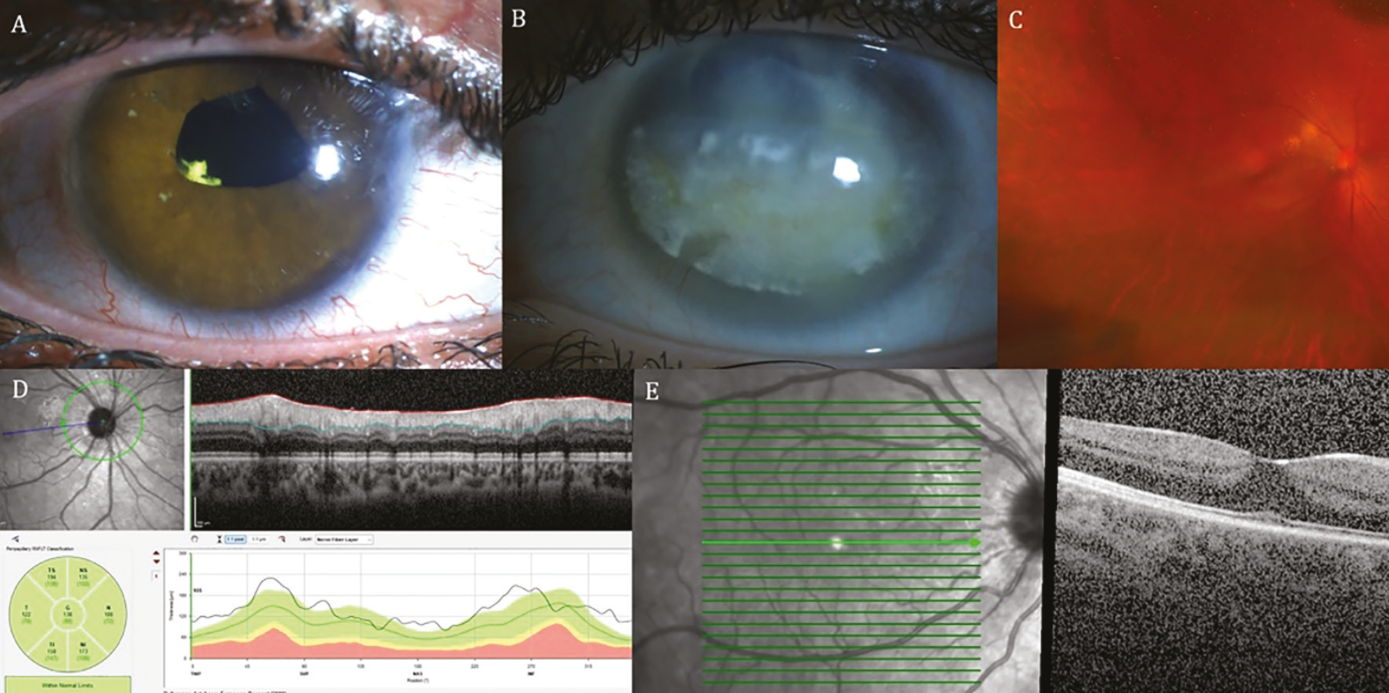
A 21-year-old woman with anti-nuclear antibody-positive juvenile idiopathic arthritis-associated uveitis presented with uncontrolled intraocular pressure (IOP) in her only seeing eye, which had undergone pars plana vitrectomy 15 years earlier for vitreous opacity and tractional retinal detachment. The fellow eye had lost vision due to chronic uveitic complications and dense band keratopathy (). At presentation, the best-corrected visual acuity (BCVA) was 0.8 in the right eye, and IOP measured 36 mmHg despite maximal tolerated medical therapy. The cornea was clear and the intraocular lens was centered (). Fundus examination revealed a healthy optic disc and macula (), and peripapillary retinal nerve fiber layer and macular optical coherence tomography (OCT) were within normal limits (, ).
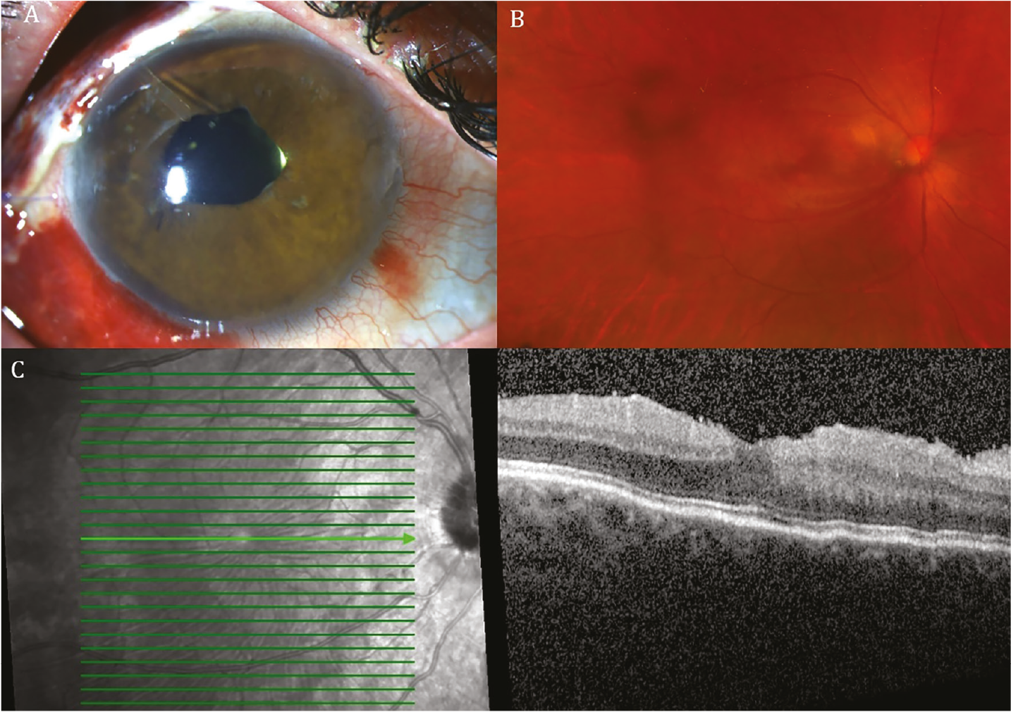
Given the patient’s monocular status and refractory IOP elevation despite maximal medical therapy, a Paul Glaucoma Implant (Advanced Ophthalmic Innovations Pte Ltd, Singapore, Republic of Singapore) was placed with an intraluminal 6-0 polypropylene ripcord (Prolene suture; Ethicon, Raritan, NJ, USA). The tube was advanced entirely within a scleral tunnel beneath a scleral flap and subsequently introduced into the anterior chamber through a 26-gauge needle tract. On postoperative day 1, IOP was 13 mmHg with stable visual acuity. However, by postoperative day 10, the patient developed acute visual deterioration to 0.1 accompanied by hypotony (IOP 3 mmHg). The anterior chamber remained well formed and tube position was appropriate (), but fundus examination revealed chorioretinal folds consistent with hypotony maculopathy (), which was confirmed on macular OCT (). Anterior chamber reformation with a 3.0% sodium hyaluronate ophthalmic viscosurgical device (Hyotek 3%; Teknomek Medikal Malzemeleri Sanayi ve Ticaret Ltd. Şti., Ergene, Tekirdağ, Türkiye) resulted in transient IOP recovery; however, recurrent hypotony developed at the first month of follow-up, necessitating further flow-modulating intervention.
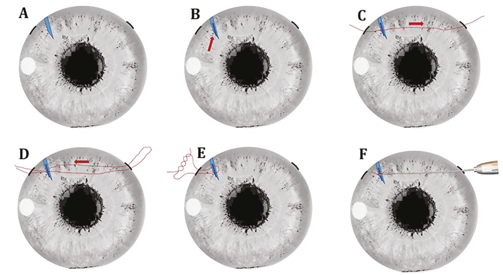
Hypotony maculopathy is a vision-threatening complication that may lead to permanent structural distortion and irreversible visual loss if not promptly addressed. To modulate aqueous outflow in a controlled and reversible manner, an ab interno, stepwise tube ligation technique was performed (-F). The intraluminal 6-0 Prolene ripcord suture introduced from the distal end of the tube during the initial surgery was left in place. For the ligation procedure, a second 6-0 Prolene ripcord suture was advanced intraluminally from the proximal end of the tube to provide partial flow restriction without complete occlusion (, ). Subsequently, a 10-0 nylon suture was advanced ab interno from the anterior surface of the tube, guided across the anterior chamber, and externalized through a contralateral paracentesis (). The externalized suture was then reintroduced into the anterior chamber and redirected posterior to the tube, creating a loop encircling the tube body (). This loop was tied externally using a 3-1-1 knot configuration and seated onto the tube body to achieve external ligation without conjunctival dissection (). Finally, the intrachamber ends of the 10-0 nylon suture were trimmed with microsurgical forceps, ensuring that the cut ends remained posterior to the tube to minimize the risk of postoperative corneal endothelial trauma (). This configuration enabled graded and reversible modulation of aqueous outflow while preserving the structural integrity of the drainage device. However, IOP remained 5-6 mmHg, accompanied by corneal edema and anterior chamber inflammation one week after the tube ligation (), suggesting that hypotony was driven not only by overfiltration but also by ciliary body hyposecretion related to uveitic flare.
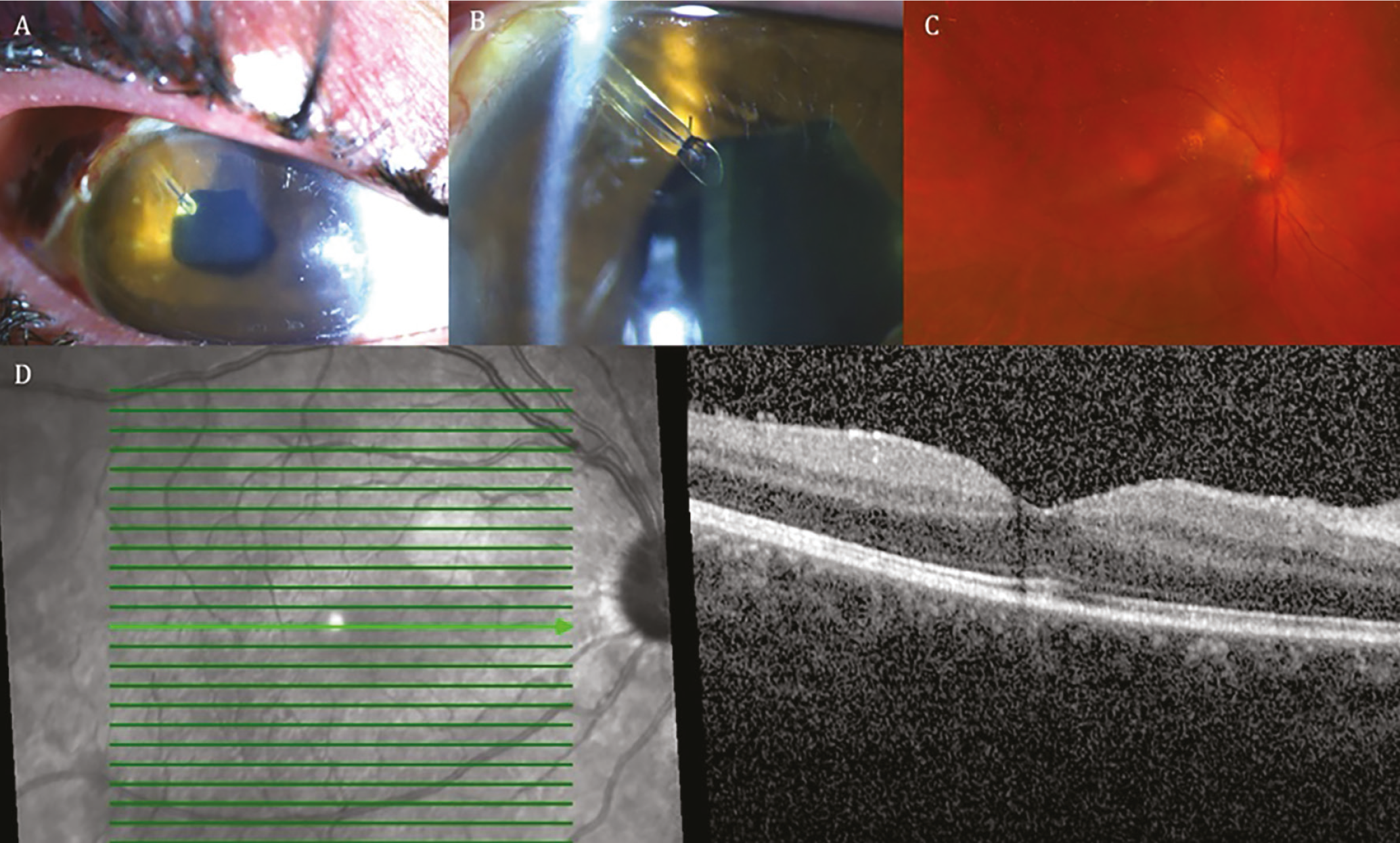
Four-quadrant anterior sub-Tenon triamcinolone injections (Kenacort-A 40 mg/mL; Bristol-Myers Squibb, New York, NY, USA) were administered to rapidly suppress inflammation and restore ciliary body function, a well-established therapeutic strategy in inflammatory hypotony., Within one week, corneal edema and intraocular inflammation resolved (), and IOP gradually increased to physiologic levels. Two months later, IOP stabilized at 16 mmHg. Fundus examination showed resolution of macular folds (), and macular OCT confirmed restoration of normal foveal contour and retinal layering (). Final BCVA returned to 0.8. The patient remained under systemic and topical anti-inflammatory/immunomodulatory treatment consisting of topical prednisolone acetate 1% three times daily (Pred Forte; AbbVie Inc., North Chicago, IL, USA), mycophenolate mofetil 500 mg twice daily (CellCept; F. Hoffmann-La Roche Ltd., Basel, Switzerland), oral methylprednisolone 32 mg once daily (Prednol; Mustafa Nevzat İlaç Sanayii A.Ş., Istanbul, Türkiye), azathioprine 50 mg twice daily (Imuran; GlaxoSmithKline, Brentford, UK), and adalimumab 40 mg once weekly (Humira; AbbVie Inc., North Chicago, IL, USA).
This case demonstrates that postoperative hypotony following glaucoma drainage device implantation in uveitic eyes should not be attributed solely to excessive aqueous outflow. Inflammatory ciliary body suppression must be considered, as its management differs fundamentally. In this patient, we implemented an ab interno, stepwise, and reversible flow-modulation strategy combining intraluminal 6-0 Prolene ripcord placement with loop-based 10-0 nylon tube ligation. This configuration not only allowed controlled elevation of IOP while inflammation was concurrently treated with sub-Tenon corticosteroid injections, but also preserved future flexibility of aqueous outflow. Should IOP rise during follow-up, graded restoration of tube patency can be achieved by first removing the intraluminal Prolene ripcord, creating an approximate 70-µm lumen, and, if further flow is required, by cutting the tube distal to the nylon ligation to establish a wider opening of approximately 127 µm. This stepwise outflow restoration preserves implant integrity, avoids surgical revision, and provides a temporal window for recovery of ciliary body function. In the present case, the initial intraluminal 6-0 Prolene ripcord was not removed during follow-up, as IOP stabilized after inflammation control and tube ligation. Nevertheless, an additional technical consideration is that removal of a 6-0 Prolene ripcord cut flush with the tube tip may be technically challenging if intraluminal adhesion develops over time. Should removal prove difficult, stepwise flow restoration may alternatively be achieved through distal tube trimming or selective release of external ligation sutures, thereby preserving implant integrity without conjunctival dissection.
From a pathophysiological standpoint, the second episode of hypotony occurring at one month was unlikely to be solely attributable to excessive aqueous outflow.
The transient IOP improvement after the initial viscoelastic injection may have coincided with temporary fluctuations in uveitic activity rather than representing definitive correction of overfiltration. Moreover, the persistence of hypotony despite mechanical flow restriction suggests that inflammatory ciliary body hyposecretion played a substantial role. The rapid anatomical and pressure recovery following sub-Tenon corticosteroid administration further supports this interpretation and underscores the importance of aggressive inflammation control in similar cases.
Although small-bore tube design and intraluminal ripcord stenting are intended to mitigate early postoperative hypotony, such measures provide partial rather than complete occlusion. Residual low-grade outflow may persist around the stent, particularly before capsule maturation increases resistance. In uveitic eyes, postoperative inflammatory fluctuations may further suppress ciliary body function, thereby shifting the balance toward hypotony despite flow-restricting strategies. Taken together, the prolonged and recurrent hypotony observed in this case most likely reflected a multifactorial process in which modest residual outflow and inflammation-related hyposecretion acted synergistically.
A further potential mechanism to consider in cases of persistent hypotony after drainage device implantation is peritubular leakage. In this patient, the tube was advanced entirely within a scleral tunnel and covered by a scleral flap without secondary scleral surface entry, reducing the likelihood of clinically significant leakage. The anterior chamber remained well formed and no Seidel-positive leakage was detected. Nevertheless, subtle microleakage cannot be completely excluded in prolonged hypotony scenarios.
In conclusion, early recognition of inflammatory hypotony, combined with targeted anti-inflammatory therapy and a flexible, reversible approach to aqueous outflow modulation, may enable both anatomical and functional recovery, even in monocular patients in whom visual preservation is critical. While this report represents a single case and should therefore be considered a proof-of-concept experience, it highlights a potentially adaptable strategy for managing complex hypotony scenarios. Further evaluation in larger case series is warranted to assess reproducibility, safety, and long-term outcomes.
Footnotes
Ethics
Informed Consent: Written informed consent was obtained from the patient for the publication of their clinical information and images.
Authorship Contributions
Surgical and Medical Practices: A.Y.Ü., İ.G.G., H.B.Ö., Concept: A.Y.Ü., İ.G.G., Design: A.Y.Ü., İ.G.G., Data Collection or Processing: A.Y.Ü., İ.G.G., B.U., B.A., Analysis or Interpretation: A.Y.Ü., İ.G.G., Literature Search: A.Y.Ü., Writing: A.Y.Ü.
Conflict of Interest: No conflict of interest was declared by the authors.
Financial Disclosure: The authors declared that this study received no financial support.
References
Škrlová E, Svozílková P, Heissigerová J, Fichtl M. Pathogenesis and current methods of treatment of secondary uveitic glaucoma. A review. Cesk Slov Oftalmol. 2023;79:111-115.
Kalogeropoulos D, Sung VC. Pathogenesis of uveitic glaucoma. J Curr Glaucoma Pract. 2018;12:125-138.
Chaku M, Bajwa A, Lee JK, Netland PA. Treatment of uveitis and outcomes of glaucoma drainage implant surgery: a meta-analysis. Ocul Immunol Inflamm. 2020;28:833-838.
Satana B, Yalvac IS, Sungur G, Eksioglu U, Basarir B, Altan C, Duman S. Ahmed glaucoma valve implantation for uveitic glaucoma secondary to Behçet disease. J Glaucoma. 2015;24:607-612.
de Smet MD, Gunning F, Feenstra R. The surgical management of chronic hypotony due to uveitis. Eye (Lond). 2005;19:60-64.
Thomas M, Vajaranant TS, Aref AA. Hypotony maculopathy: clinical presentation and therapeutic methods. Ophthalmol Ther. 2015;4:79-88.
Ferreira BG, Marinho DR, Diligenti FT. Effects of sub-Tenon’s triamcinolone injections in patients with uveitis. Arq Bras Oftalmol. 2018;81:323-329.
Nilforushan N, Abolfathzadeh N, Najafzadeh F, Samimifar D. Subtenon triamcinolone injection to treat serous choroidal detachment after glaucoma surgery: a case series. Eur J Ophthalmol. 2023;33:319-323.